Services
Our Services
-
comprehensive eye examination
-
cataract assessment & Microincision Surgery
-
computerised testing for glasses
-
Glaucoma Care
-
pediatric eye care
-
retina checkup
-
diabetic retinopathy screening
-
intra ocular (Eye) pressure testing
-
retinopathy of prematurity screening
-
amblyopia therapy / lazy eye therapy
-
treatment for dryness, allergies, infection of eye
-
Phacoemulsification
-
Small incision cataract surgery (SICS)
-
Pterygium excision
-
Pediatric cataract
-
Pediatric oculoplasty
-
Squint
-
Phaco-trabeculectomy
Visual Field Test
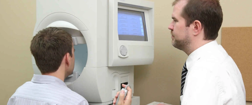
A visual field test is an essential tool that helps measure how well you see in all directions—up, down, left, right, and straight ahead—without moving your eyes. Known medically as a perimetry test, it plays a major role in detecting and monitoring eye and neurological conditions such as glaucoma.
While not always included in every routine eye exam, visual field testing is commonly performed when symptoms, risk factors, or existing conditions are present. It helps detect subtle changes in your side (peripheral) vision and provides detailed insight into how your entire visual system is functioning.
Components of Visual Field Test
- Extent of Vision Without Eye Movement: Measures how far you can see in all directions while looking straight ahead. Helps identify reductions in peripheral vision, even before you notice any changes.
- Sensitivity to Varying Light Intensities: Tests how well different areas of your vision detect lights of varying brightness—not just dim lights. Identifies specific spots where vision may be weaker or less responsive.
Types of Visual Field Tests
- Amsler Grid: A simple screening tool using a grid pattern to detect issues in central vision. It’s not a full perimetry test but can highlight signs of macular disease.
- Confrontation Test: A basic gross screening method where the provider uses their fingers or a small object to check peripheral vision while you look straight ahead.
- Automated Machine Tests: More precise and widely used in clinical practice:
- Humphrey Visual Field Test
- Octopus Perimeter
- Goldmann Perimeter
- Insight into Nervous System Health: The test can detect signs of dysfunction in the retina, optic nerve, or brain. Certain conditions show classic patterns—for example, pituitary tumors often cause a pattern of vision loss known as bitemporal hemianopia (loss of outer visual fields in both eyes).
- Used in Follow-Up and Monitoring: The test may be repeated at follow-up appointments—weeks or months later—not typically "right away" unless accuracy is questioned. Repeated testing tracks disease progression and treatment effectiveness.
Conditions Monitored or Evaluated
- Glaucoma – Detects early and progressive loss of peripheral vision.
- Macular Degeneration – Although not diagnosed primarily through this test, it can reveal central field defects linked to the condition.
- Stroke or Multiple Sclerosis (MS) – Can detect patterns of visual field loss caused by these neurological conditions.
- Pituitary Gland Tumors – Often cause a specific pattern of peripheral vision loss, helpful in diagnosis and monitoring.
Benefits of Visual Field Test
- Early Detection of Visual Changes
- Finds changes in vision before symptoms are noticeable, especially in conditions like glaucoma.
- Helps catch problems that may not be visible during a standard eye exam. - Monitors Treatment Success
- Shows whether a disease is stable, improving, or worsening.
- Allows your provider to adjust treatment plans based on detailed visual feedback. - Non-Invasive and Comfortable
- No injections, eye drops, or physical contact required.
- Completely painless and requires no special preparation. - Supports Informed Diagnosis
- Provides insights about the eye and brain’s visual pathways.
- Helps rule out or support neurological and structural issues. - Doctor-Guided Testing Frequency
- Testing intervals are set by your eye care provider based on your condition.
- Some people may need testing every few months, while others may only need it annually. - Educational Tool in Care
- Your provider uses the results to help explain changes in your vision.
- Promotes understanding and helps guide discussions about future treatment options. - Simple Yet Powerful:
- Though the test is easy to take, it reveals vital information.
- Can significantly impact early diagnosis and protect long-term vision health.